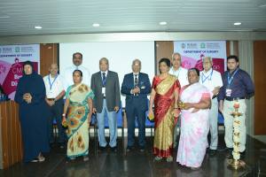
The CME on Breast Diseases was organised by the Department of General Surgery of Mahatma Gandhi Medical College. It was inaugurated by Prof.V.Sethuraman, Vice-Chancellor, Sri Balaji Vidyapeeth in the presence of Prof.M.Ravishankar, Dean, MGMCRI, Prof.V. Nirmal Coumare, medical Supdt., Prof.N.Ananthakrishnan, Dean – Research, MGMC RI, Prof.Robinson Smile and Prof.M.Ramanathan, Head, Department of Surgery
This program was very unique in that it was inaugurated by the breast cancer survivors and the lighting of the Kuthuvilaku was done by them. There was a rendition on the song about Cancer survival. Moreover, a movie on Cancer Survival was also screened. There were about 150 delegates from Pondicherry and Chennai who took part in this meeting
Emr Prof.Raghavan Narasimhan gave an overview of basic surgical pathology in breast diseases.
Dr. V.C. Sunitha, Consultant in the Department of Radiodiagnosis, MER enlightened us about the role of imaging modalities in Breast diseases
Dr. M.Prema, Consultant Breast Surgeon , Rajah Muthiah Medical College, Chidambaram elaborately discussed about benign breast diseases , which constitute about 90% of the patient population coming with breast complaints. Broadly the questions to be met are- whether it is a cancer? Whether to intervene or not? If yes , how to intervene ? What is the medical management ? What are the surgical options available? What is the extent of surgery? Whether it can become cancerous? What are the follow up protocols?
In this light, she went on to discuss the clinical classification of benign breast diseases which fall under the categories as congenital/ developmental anomalies, infectious/ inflammatory disorders, proliferative/ tumor like lesions and the miscellaneous category. The pathogenesis of aberations in the normal development and involution of the breast was discussed with more details on the influence of the various hormones on the breast. Enough stress was laid on the importance of detailed history taking and clinical examination. The major complaints that patients come with are mastalgia, physiological tenderness/ swelling, nodularity/ lumpiness, dominant mass, nipple discharge, nipple changes, breast infections.
Since we are in an era of defensive medicine radiological assessment and triple assessment take a prime place. Pathological assessment plays a vital role for both documentation and for further follow ups. The various methods of taking tissue biopsies namely core needle biopsy, excision and incision biopsy along with newer techniques like vaccum assisted biopsies, stereotactic biopsy, wire localisation, radio occult lesion localisation and biopsy were discussed. Mastalgia, fibroadenoma, simple cyst, fibrocystic dissease, duct papilloma, duct ectasia, inverted nipple, premature thelarche, lactational and non lactational breast abscess, mastitis, gynecomastia was done. Concluding, she emphasized on ruling out malignancy by triple assessment and the need for appropriate risk stratification of benign lesions .
Dr.Selvi Radhakrishna, Consultant Oncoplastic Surgeon, Chennai Breast centre, Chennai gave a lecture on triple assessment and management of early breast cancer. She started off with a case scenario of a 44 year old woman with a two weeks complaint of breast pain , she also noticed a thickening in her breast. With this as background, the detailed relevant history to suspect or rule out malignancy was discussed. In such cases, history of the duration of the complaint, associated pain, discharge, menarche, menstrual history, parity, age at first child birth , breast feeding history, contraceptives , Hormone replacement therapy, other medications, family history of breast and related malignancies have to be probed. After history and examination follows the radiological and pathological assessment. For patients, more than 35 years of age mammogram is advised and for patients, less than 35 years of age ultrasound followed by mammogram only if warranted is advised . Mammogram and ultrasound findings that suggest malignancy and their significance was described in detail. In her view she favours core biopsy over a fine needle aspiration and cytology as fnac has the following disadvantages – it may turn out to be inconclusive or inadequate, higher chance of false positive or false negative results, the disappearance of tumor post Neoadjuvant treatment leaving no tissue to check for receptor status. On the other hand, core biopsy is more accurate, giving receptor status to plan for NACT, and also allows for tumor banking. Early breast cases are ideal for breast conservation with an appropriate patient selection. She gave an overview of Oncoplastic techniques involving volume displacement and volume replacement techniques. In early breast cancer, mastectomy is done if it is the patients choice due to logistic reasons, if there is extensive disease, if it was a multicentric disease, if the patient cannot comply or afford radiation treatment. She mentioned the two commonly used techniques of Sentinel lymph node biopsy. The surgery to be accompanied by chemotherapy, radiotherapy, hormonal therapy along with anti -Her 2 treatment appropriately. She concluded with a video demonstration of a modified radical mastectomy using a smilie incision with level three axillary clearance.
Dr. Kadambari Dharanipragada, Professor of Surgery, Head, Medical education, JIPMER delivered a talk on locally advanced breast cancer. Breast malignancies over the size of 5 cm (T3,T4) with fixed ipsilateral axillary nodes, metastasis to infra clavicular, supraclavicular or internal mammary nodes (N3,N4) fall under this category. Here too enough emphasis had been laid on the history , clinical examination, imaging and pathological examination. It is important to work up extensively for metastatic disease in these patients. Hence apart from the routine investigations, CECT thorax , abdomen , bone scan and FDG PET-CT scan play a major part. Treatment approach is multimodal therapy . Neoadjuvant chemotherapy with taxanes, anthracyclins are given for 6 to 8 cycles and for patients with triple negative disease platinum based chemo therapy is preferred. Along with chemotherapy anti HER 2 receptor agents like trastuzumab, lapatinib, pertuzumab are used. Hormonal therapy targeting estrogen receptors such as tamoxifen, letrazole, anastrazole are advocated in estrogen receptor positive patients. Also a mention of the newer agents for systemic neoadjuvant therapy namely everolimus, palbociclib, vorinostat was made. The response to the neoadjuvant therapy is made by three methods- clinical examination using WHO criteria, imaging which uses RECIST criteria, pathology using AJCC and other systems. These were elaborated in detail, highlighting the various types of responses on MRI to the chemotherapy. The various methods of MRI imaging was mentioned briefly. Response evaluation methods and residual disease assessment is done by AJCC criteria, residual cancer burden index, miller payne method, sataloff classification. Where ever appropriate PET- CT has to be done to assess the disease burden in doubtful cases. Once Neoadjuvant chemotherapy is completed and response assessed the other modalities also need to be implemented namely surgery , radiotherapy, hormonal therapy and anti Her 2 therapy. Surgery for these cases can be breast conservation surgery, modified radical mastectomy, toilet mastectomy based on the response and disease progression. On completion of treatment these patients need to be followed every 3-12 months with history and clinical examination, annual mammograms, metastases work up only if sympyomatic, gynaecological screening for patients on tamoxifen, Bone densitu monitoring for patients on aromatase inhibitors